


OCT Eye Exams in Edmonton
Let’s Take a Deeper Look Into Your Eye Health
What is 3D OCT Imaging?
OCT stands for Optical Coherence Tomography, and it is one of the most valuable tools we have for understanding what is happening inside your eye.
Think of it like an MRI for your eyes. In just a few seconds, it captures high-resolution, cross-sectional images of your retina, optic nerve, cornea, and iris. This gives us a detailed, three-dimensional view of your eye’s internal structures. The test is completely non-invasive, does not touch the eye, and is quick and comfortable.
What makes OCT so powerful is what it can reveal. Many serious eye conditions, including glaucoma, macular degeneration, and diabetic eye disease, often develop without noticeable symptoms. OCT allows us to detect subtle changes in the layers of your eye before they affect your vision, giving us the best opportunity to intervene early.
At Helio Optometry, OCT imaging is a routine part of our comprehensive eye exams. We believe thorough eye care goes beyond simply checking your prescription.

Why Do We Use OCT Imaging?
A standard eye exam gives us a lot of information, but it has limits. OCT imaging allows us to see through the retina and evaluate the deeper layers of the eye that would otherwise be invisible, giving us a far more complete picture of your eye health.
This technology has become the gold standard in eye care for good reason. It provides critical insight into conditions like glaucoma, age-related macular degeneration, diabetic retinopathy, and a range of other retinal disorders, often detecting changes before any symptoms appear.
The level of detail it captures is remarkable. The retina is no thicker than 0.5mm, and our Nidek 3000 OCT can scan down to just 4 microns (0.004mm). That level of precision gives our optometrists a detailed, layered view of your eye structures that simply isn't possible any other way.
OCT imaging is also essential for specific patient groups. It's the preferred tool for monitoring patients on Plaquenil (Hydroxychloroquine), a medication commonly used to treat malaria and arthritis that can affect the retina over time. For patients with a family history of glaucoma, it allows us to detect early or subtle changes in the optic nerve, opening the door to earlier treatment or simply the reassurance that things are stable.
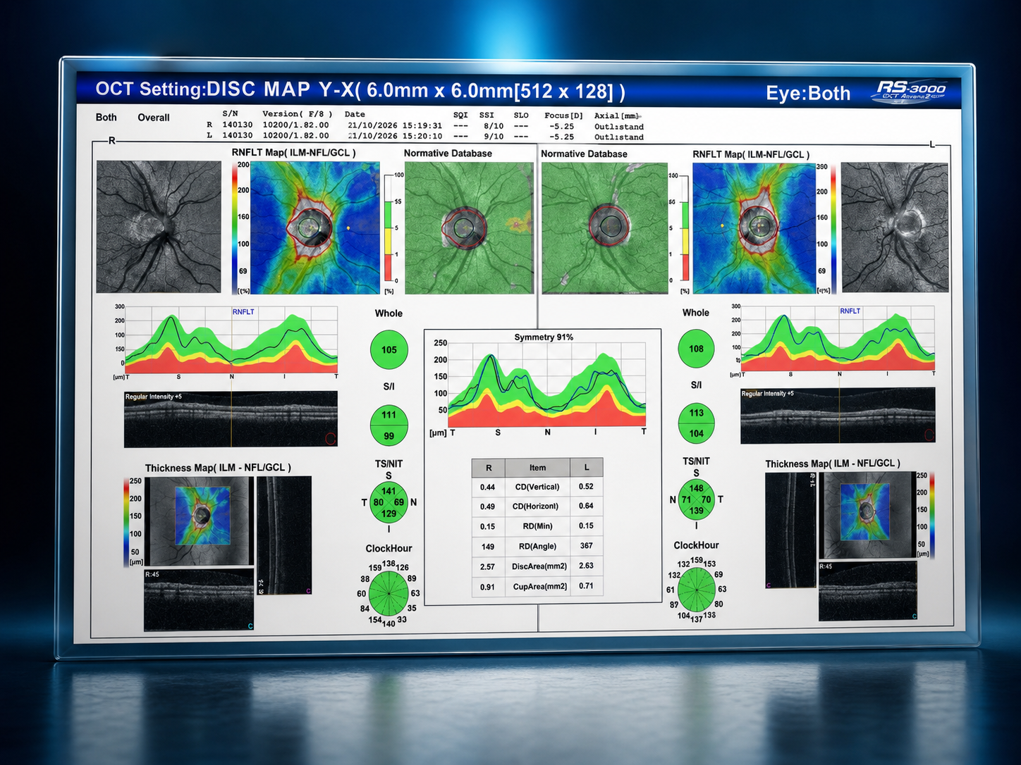
What Does an OCT Scan Actually Show?
An OCT scan gives us a detailed, cross-sectional view of the structures at the back of your eye. It allows us to see far more than what is visible during a standard eye exam.
We use it to measure the thickness of the retinal nerve fibre layer (RNFL) and evaluate the optic nerve for early signs of glaucoma. It also maps the macula, helping us detect subtle changes related to macular degeneration or diabetic eye disease.
OCT lets us compare both eyes, assess symmetry, and track changes over time. We can analyze important measurements like the cup-to-disc ratio and retinal thickness across different regions.
Perhaps most importantly, it allows us to visualize the individual layers of the retina. This helps us identify swelling, thinning, or damage long before it begins to affect your vision.
It is one of the most powerful tools we use to detect problems early and monitor your eye health with precision.
Why Do We Use OCT Imaging?
A standard eye exam gives us a lot of information, but it has limits. OCT imaging allows us to see through the retina and evaluate the deeper layers of the eye that would otherwise be invisible, giving us a far more complete picture of your eye health.
This technology has become the gold standard in eye care for good reason. It provides critical insight into conditions like glaucoma, age-related macular degeneration, diabetic retinopathy, and a range of other retinal disorders, often detecting changes before any symptoms appear.
The level of detail it captures is remarkable. The retina is no thicker than 0.5mm, and our Nidek 3000 OCT can scan down to just 4 microns (0.004mm). That level of precision gives our optometrists a detailed, layered view of your eye structures that simply isn't possible any other way.
OCT imaging is also essential for specific patient groups. It's the preferred tool for monitoring patients on Plaquenil (Hydroxychloroquine), a medication commonly used to treat malaria and arthritis that can affect the retina over time. For patients with a family history of glaucoma, it allows us to detect early or subtle changes in the optic nerve, opening the door to earlier treatment or simply the reassurance that things are stable.
Your OCT Imaging Questions,
Answered
-
Not at all. OCT is completely non-invasive and requires no contact with your eye. You simply rest your chin on a support, look at a target light, and the scan is done in a matter of seconds. Most patients barely notice it.
-
A standard eye exam evaluates how well you see and checks the visible structures of your eye. OCT goes much further, capturing detailed cross-sectional images of the retina, optic nerve, and other internal structures that can't be seen with the naked eye alone. It's the difference between looking at the surface and seeing what's underneath.
-
For most healthy adults, once a year alongside your comprehensive eye exam is sufficient. If you have a condition like glaucoma, macular degeneration, or diabetes, or if you take medications like Plaquenil, your optometrist may recommend more frequent scans to monitor any changes over time.
-
OCT imaging is covered by Alberta Health Care when it is medically necessary. However, it is not billed routinely as part of a standard eye exam. Alberta Health Care also limits the number of covered scans within a given year, so if additional scans are required beyond that, there may be out-of-pocket costs. Our team will always walk you through what's covered before your exam so there are no surprises.
-
Yes, OCT imaging is considered safe during pregnancy. The scan uses light waves rather than radiation, so there is no known risk to you or your baby. It's worth noting that pregnancy can cause temporary changes in the eyes, including shifts in prescription. Pregnancy can also increase the risk of developing gestational diabetes, and for those already living with Type 1 or Type 2 diabetes, it can accelerate the progression of diabetic retinopathy. For these reasons, staying on top of your eye health during pregnancy is especially important. As always, let your optometrist know you are pregnant so they can take your full health picture into account.
-
OCT is one of the most powerful tools we have for detecting early signs of glaucoma, but it is one piece of a larger puzzle. Glaucoma diagnosis relies on several factors together, including OCT scans of the optic nerve and retinal nerve fibre layer, eye pressure measurements, visual field testing, and a thorough clinical examination. OCT is particularly valuable because it can detect structural changes in the optic nerve before vision loss begins, often years before a patient would notice anything wrong. So while OCT alone cannot diagnose glaucoma, it plays a central role in catching it early and monitoring it over time.

Is It Time to Take a Closer Look At Your Eye Health?
Don't wait for symptoms to tell you something is wrong. Many serious eye conditions develop silently, and OCT imaging is one of the best ways to catch them early.
Visit Us in Edmonton —>